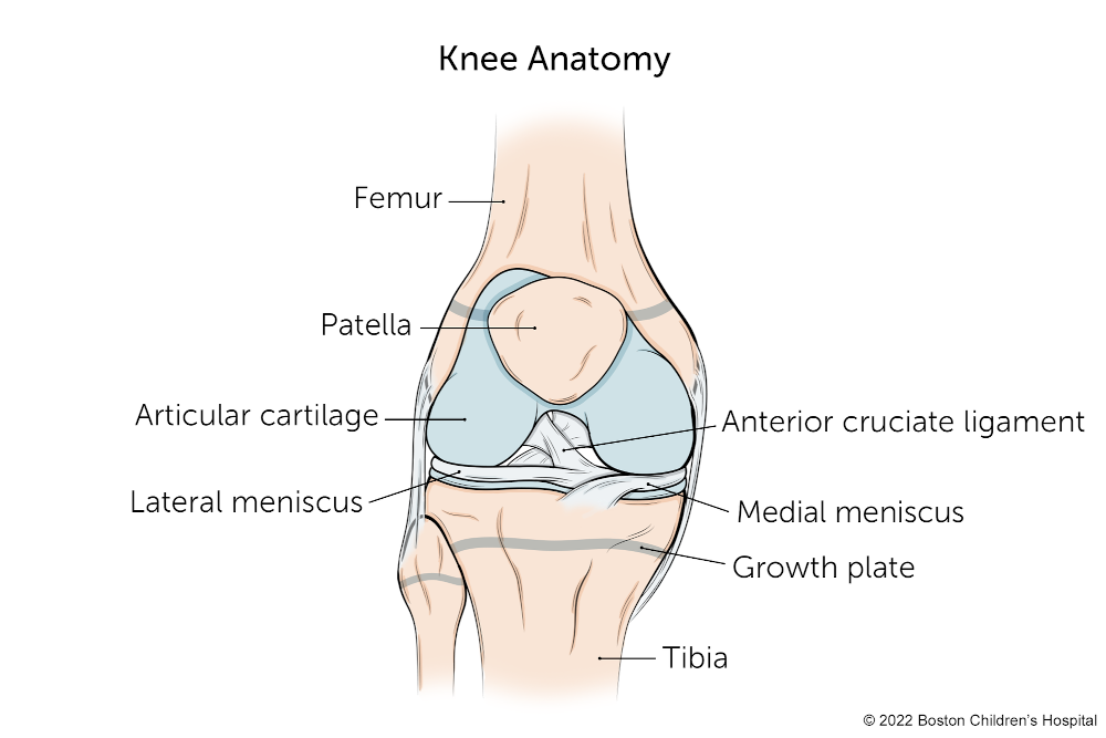
Three different types of cartilage are found in the body:
- articular or hyaline cartilage: covers joint surfaces
- fibrocartilage: such as in the knee meniscus and vertebral disk
- elastic cartilage: such as the outer ear
Articular cartilage is a complex, living tissue that lines the bony surface of joints. It provides a low-friction surface, enabling the joint to withstand weight-bearing movements, both for daily activities as well as athletics, including walking and stair climbing, and work-related activities. In other words, articular cartilage is a very thin shock absorber.
Articular cartilage injuries can occur as a result of either traumatic mechanical destruction or progressive mechanical degeneration (wear and tear).
Mechanical destruction of articular cartilage
With mechanical destruction, a direct blow or other trauma can injure the articular cartilage.
- Because articular cartilage has no direct blood supply, it has little or no capacity to repair itself from mechanical destruction.
- Depending on the extent of the damage and location of the injury, it is sometimes possible for the articular cartilage cells to heal.
- If the injury penetrates the bone beneath the cartilage, the underlying bone provides some blood to the area, improving the rate of healing.
Occasionally, an articular cartilage fragment completely breaks loose from the underlying bone. This chip, called a loose body, may float in the joint, interfering with normal joint motion.
Mechanical degeneration of articular cartilage
Mechanical degeneration (wear and tear) of articular cartilage occurs with the progressive loss of the normal cartilage structure and function.
- This initial loss begins with cartilage softening, then proceeds to fragmentation.
- As the loss of the articular cartilage lining continues, the underlying bone has no protection from the normal wear and tear of daily living and begins to break down, leading to osteoarthritis.
Also known as degenerative joint disease, osteoarthritis is characterized by three processes:
- a progressive loss of cartilage
- the body's attempted to repair the cartilage
- the destruction of the bone underneath the articular cartilage
The cause of osteoarthritis is poorly understood, but lifelong moderate use of normal joints does not increase the risk. Factors such as high-impact twisting injuries, abnormal joint anatomy, joint instability, inadequate muscle strength or endurance, and medical or genetic factors can contribute to osteoarthritis.
What are the symptoms of an articular cartilage injury?
- Knee swelling and vague pain. At this point continued activity may not be possible.
- If a loose body is present, words such as “locking” or “catching” might be used to describe the problem.
- With mechanical degeneration (wear and tear), the patient often experiences stiffness, decreased range of motion, joint pain, and/or swelling.
How does Boston Children's Hospital approach articular cartilage injuries?
Depending on the severity of your child's articular cartilage injury, treatment may be surgical or non-surgical. At Boston Children's, doctors are committed to repairing your child's knee in the least invasive manner possible, including physical therapy and tips on lifestyle changes. Surgery is only used in the most severe cases of articular cartilage injuries.
Articular Cartilage Injury | Diagnosis & Treatments
How does a doctor know my child has an articular cartilage injury?
The physician examines your child's joint, looking for:
- decreased range of motion
- pain along the joint line
- swelling
- fluid on the knee
- abnormal ailment of the bones making up the joint
- ligament or meniscal injury
Because an articular cartilage injury is hard to diagnose, your child's doctor may also require:
- Magnetic resonance imaging (MRI): A diagnostic procedure that uses a combination of large magnets, radio frequencies, and a computer to produce detailed images of organs and structures within the body
- Arthroscopy: A minimally invasive outpatient procedure that inserts a small camera into the joint for the doctor to inspect.
How we treat an articular cartilage injury
When a joint is injured, the body releases enzymes that may further break down the already damaged articular cartilage.
- Injuries to the cartilage that do not extend to the bone generally do not heal on their own.
- Injuries that penetrate to the bone may heal, but the type of cartilage that is laid down is structurally unorganized and does not function as well as the original articular cartilage.
Defects smaller than two centimeters have the best prognosis and treatment options. Those options include arthroscopic surgery, which can remove damaged cartilage and increase blood flow from the underlying bone (e.g. drilling, pick procedure). For larger defects, it may be necessary to transplant cartilage from other areas of the knee (joint).
Non-surgical options
For patients with osteoarthritis:
- physical therapy
- lifestyle modification (e.g. reducing activity)
- bracing
- supportive devices
- oral and injection drugs (i.e. non-steroidal anti-inflammatory drugs, cartilage protective drugs)
- medical management
Surgical options
Surgery often only provide short-term relief, and depends on the severity of the osteoarthritis:
- Tibial or femoral osteotomics (cutting the bone to rebalance joint wear) may reduce symptoms, help to maintain an active lifestyle, and delay the need for total joint replacement.
- Total joint replacement can provide relief for the symptom of advanced osteoarthritis, but generally requires changes in a patient's lifestyle and/or activity level.